Everything you need to know about heel spur: what is it, cause, symptoms, general practitioner, treatment, surgery and exercises
More and more people are suffering from heel spur. It is about pain at the location where the foot sole tendon is attached to the heel bone. This is a thick and wide band of rigid tissue that runs under the foot and is attached to the heel bone. Upon heel spur, a sharp pain may occur in the heel under the foot when you put strain on it. In most cases, the pain usually spontaneously disappears within four months. The medical name for this condition is ‘calcaneus spur’.
What is heel spur exactly?
The main culprit is calcium deposition at the place where the foot sole is attached to the heel bone. Overload can cause small cracks in the tendons. By calcium deposition, the body tries to reinforce the attachment of the foot sole to the heel bone. This calcium deposit is called heel spur. This hook on the heel bone can be clearly observed on an X-ray. An estimated 30 to 75 percent of the grown-ups has heel spur, which however does not need to result in pain.
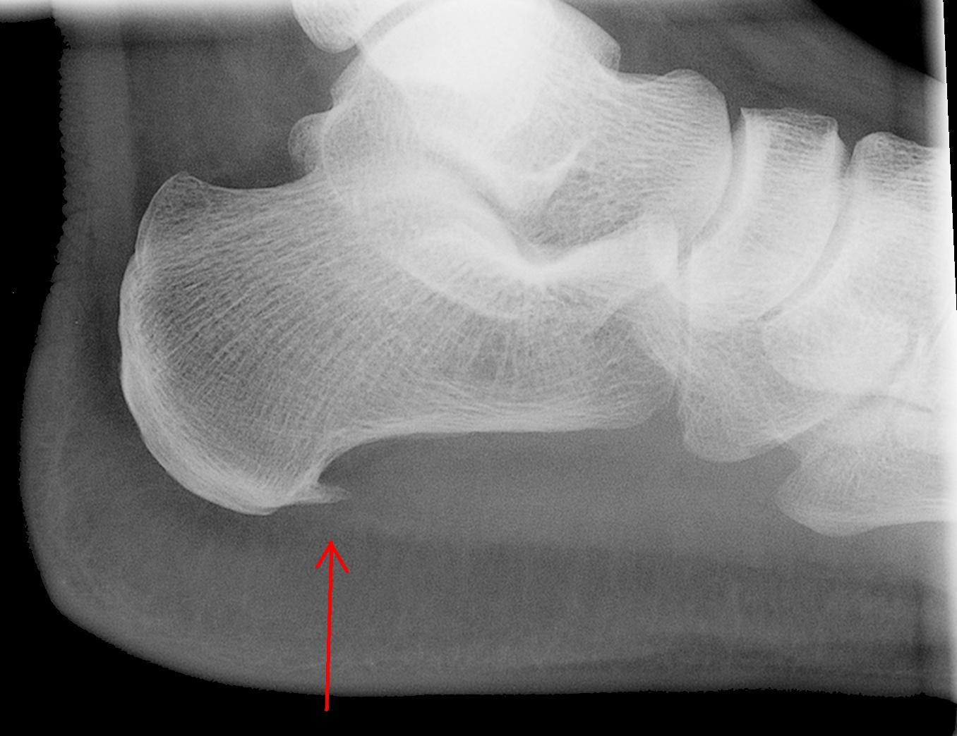
Heel spur is easy to recognize on an X-ray
What is the cause of a heel spur?
Heel spur is a relatively ‘modern’ condition. Twenty years ago, this type of heel pain was not as frequent as today. It is assumed that the increase in this form of overload injury is due to the increased popularity of sports. For example, running on a hard surface can irritate the tendon attachments to the heel. But athletes who have to run a lot and jump – like tennis players and volley ballers – are at risk of developing heel spur. This also applies to intensive hikers. Overweight, wrong walking pattern and wrong shoes can also lead to heel spur. In people who perform a standing profession, this disorder is often inevitable. Obviously, it’s called ‘Policeman’s Heel’ in English. This condition is most common in people between the ages of 40 and 60. Women are more likely to suffer from heel spur than men.
What are the symptoms of heel spur?
The main symptom is a stinging or sore pain under the foot close to the heel. This is mainly during the morning or after a longer rest period. The pain gets less if you walk a bit. The nerves in your foot then adapt, causing the pain in the heel to disappear or decrease. The foot sole tendon can also feel tight and tense. The pain can become worse over time. Then you have to walk a little longer to get pain relief.
When should you consult a GP with heel spur?
Although an annoying disorder, it’s not dangerous one. Within one to four months the worst pain disappears naturally. The calcium deposit will still be visible on an X-ray, but do not cause any symptoms. In the early stage of heel spur it is wise to buy good shoes and watch your body posture when walking. If you are too heavy, try to lose a few pounds. Against the pain of heel spur, you can use paracetamol, anti-inflammatory analgesics or a natural alternative in the form of liquid Green-lipped mussel with Bio-Curcumin. These pain relievers do not heal the heel spur, but can provide some relief. When the symptoms persist for a long time it’s wise to make an appointment with your GP, podiatrist or physical therapist.
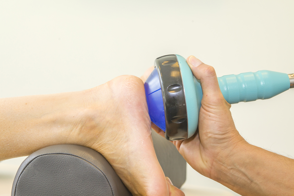
Heel spur treatment with shockwave therapy
How is heel spur treated?
The podiatrist will provide you with support soles that are completely adapted to your posture and configuration of your feet. The physiotherapist increasingly provides shockwave as a standard treatment at heel spur. He operates a device aiming sound waves at the sites where the calcium deposits are located. This improves circulation and metabolism, which reduces the pain. The GP can give an injection with corticosteroids with variable results.
When is surgery performed?
In most people, surgery is unnecessary because the heel pain automatically disappears within four months. Surgery is performed in less than 5 percent of patients with heel spur because unfortunately they are experiencing severe and persistent complaints. If you decide to do so, you should be aware of the possible complications after such surgery. For example, a weakening of the foot arch may occur. Also, nerves around the calcium deposit can be damaged. This may result in insensitivity in various places under the foot and at the heel. During surgery, the foot sole is partially detached from the heel bone. This reduces the pressure. The calcium deposit and any damaged tissues are removed.
Exercises at heel spur
Exercise 1:
Stand with your toes on a stairway or sidewalk and lower your heels. Repeat this exercise several times.
Exercise 2:
Put your hands against a wall. Move one leg forward while turning the other leg backwards. Then bend the whole body forward. Hold this position a few seconds and repeat the exercise the other way round.
Exercise 3:
With your back you are leaning against a wall. Pull your knees at a slow pace until you have taken a seat. Hold this position for a few seconds. Repeat this exercise several times.
Exercise 4:
Sit down on a chair and cross one leg across the other knee. Pull the toes backwards. Hold this position for a few seconds. Repeat this exercise several times.

Share this page
Tweet


Download for free the booklet ‘Moving without pain’ with a retail value of $6.75 / £4.95.

Any questions? Please feel free to contact us. Contact us.
Download for free the booklet ‘Moving without pain’ with a value of $6.75 / £4.95.
Do you have questions? We like to help you. Contact us