Cartilage is an important tissue in the joints, which cover the surface of both bony ends. It is stiffer and less flexible than a muscle, but not as hard as a bone. 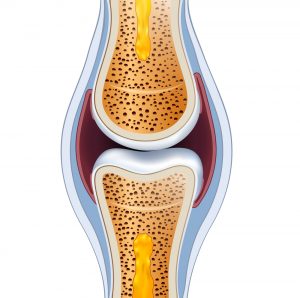
This way, the cartilage is able to carry out its primary function very well, which is decreasing the friction between the moving parts of the joints enabling them to smoothly slide among each other. This way, the surrounding tissue cannot get damaged. Cartilage needs to be strong because regularly, one single joint must carry the entire body weight. For example the knee joint when standing on one leg. Therefore, it needs to be resistant to massive forces and evenly distribute and somewhat alleviate the pressure to the joint. Preservation of healthy cartilage is therefore essential to keep the locomotion apparatus in a healthy condition. This gives you the freedom to go where ever you want.
What is the difference between hyaline and fibrous cartilage?
With regards to the locomotion apparatus there are two types of relevant cartilage. The bony ends of the joints are covered with hyaline cartilage. This is also called vitreous cartilage. It has a bluish-milky like color and is a little translucent. Hyaline cartilage is glassy enabling the joint ends to move across each other smoothly. Fibrous cartilage is white and is present at locations with large tensile load. It is about the intervertebral discs, the meniscus in the knee and the jaw joint. Also the connection between both pubic bones consists of fibrous cartilage. In osteoarthritis, the hyaline cartilage can be literally worn out to the bone. Through blood supply from the bone, sometimes fibrous cartilage is formed. This does diminish the pain, but cannot take over the role of hyaline cartilage. This process is basically no more than an ‘emergency repair’.
What is the composition of hyaline cartilage?
Hyaline cartilage consists of water for 90% making it impossible to visualize on an X-ray. However, such a photo can give the orthopedic surgeon a lot of information about the condition of the cartilage. A normal gap between both joint halves indicates that the flexible tissue is still intact. In the knee this gap needs to be 4 to 5 mm for instance. When the cartilage is partially worn out both joint halves get closer together. This is visible on an X-ray from the narrowed gap between the joints.
The hyaline cartilage is built up from four layers that gradually merge into bone. Hyaline cartilage contains less than 5% cartilage cells or chondrocytes. These construct a framework of collagen fibers. These fibers are woven together in a spiral shaped form and have different directions and mutual connection in all four layers of the cartilage. This way a strong structure is formed. The upper layer is deformable to some extend excluding damage when forces are applied. The next cartilage layers become more firm giving them a shock absorbing function. The deepest cartilage layer consists of the firmest collagen fibers. Below the ‘real’ cartilage there is another layer of calcium rich cartilage, which forms the transition to the underlying bone.
‘Entrapped’ in this ingenious network are proteoglycans. These large molecules are also produced by the cartilage cells and need to bind water. Because of this, the proteoglycans swell. This makes the cartilage more stiff, elastic, smoother and stronger. The proteoglycans itself are composed from glycosaminoglycans, among which chondroitin sulfate, keratin sulfate and hyaluronic acid. The latter substance also plays a role in the lubrication of the cartilage. Read more about this at ‘Synovial fluid’.
Proteoglycans
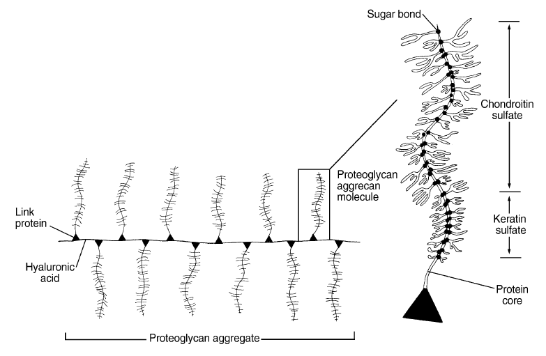
Figure: Molecular structure of proteoglycans. Proteoglycan molecules, mainly composed of chondroitin sulfate and keratan sulfate, are connected through long hyaluronic acid chains. These form the cartilage matrix by forming a tight network with each other ànd with the abundantly present collagen fibers (not shown). Figure from: Brinker, M.R. and Miller, M.D. (1999) Fundamentals of orthopedics. Philadelphia: WB Saunders; 9.
Does damaged cartilage regenerate?
Upon ageing, the quality of the cartilage might decrease. The relative content of collagen fibers will increase and the amount of water will decrease. This way the flexibility of the cartilage decreases. The consequence is that the cartilage layer is wearing off and therefore osteoarthritis develops. In addition, cartilage can also suddenly get damaged as the consequence of an accident. Small damage might eventually still get repaired. However, when the damage to the cartilage surface is a little larger, the chance for recovery is almost excluded. Then, osteoarthritis develops. There is a lot of research performed worldwide to find possibilities in order to repair damage cartilage again. At this time, there is a large clinical study going on with regard to stem cell therapy in which various European hospitals participate. At this time, the UMC Utrecht is experimenting with cartilage that can be produced by a 3D printer. Recently, one million euro in subsidy has been provided for a study with a cartilage adhesive plaster. Australian surgeons are busy developing a ‘Biopen’, with which lightly damaged cartilage can be repaired. In addition, the UMC Utrecht and the Maartens clinic in Woerden are performing research regarding the possibilities to stop the breakdown of cartilage and let it grow back. All these future treatment techniques are however in the research phase. The coming years it will become clear whether all these treatments are also practically effective. However, medical scientists agree on one thing: in time an adequate solution will be found to stop osteoarthritis!
Can damaged cartilage regenerate? Read more about solutions in osteoarthritis.

Share this page
Tweet


Download for free the booklet ‘Moving without pain’ with a retail value of $6.75 / £4.95.

Any questions? Please feel free to contact us. Contact us.
Download for free the booklet ‘Moving without pain’ with a value of $6.75 / £4.95.
Do you have questions? We like to help you. Contact us